Can We Stop Human Heart and Start It Again
Table of Contents
- Introduction
- Position
- Functions of the Human Eye
- Types of Apportionment
- Structure of the Human being Center
- Pericardium
- Structure of the Heart Wall
- Chambers of the Eye
- Blood Vessels
- Valves
- Facts about Human Center
- Important Questions about the Human Heart
Introduction to the Human Heart
The human center is one of the nigh important organs responsible for sustaining life. It is a muscular organ with four chambers. The size of the eye is the size of about a clenched fist.
The homo heart functions throughout a person's lifespan and is one of the most robust and hardest working muscles in the man body.
Besides humans, most of the other animals also possess a centre that pumps blood throughout their torso. Even invertebrates such as grasshoppers possess a heart similar pumping organ, though they do not function the same mode a human heart does.
Also Refer:Human being Circulatory System
Position of Eye in Man Body
The human being centre is located between the lungs in the thoracic cavity, slightly towards the left of the sternum (breastbone). It is derived from the embryonic mesodermal germ layer.
The Function of Heart
The part of the heart in whatsoever organism is to maintain a constant flow of blood throughout the torso. This replenishes oxygen and circulates nutrients amidst the cells and tissues.
Post-obit are the main functions of the heart:
- Ane of the primary functions of the human middle is to pump claret throughout the trunk.
- Blood delivers oxygen, hormones, glucose and other components to diverse parts of the body, including the man heart.
- The heart also ensures that acceptable blood force per unit area is maintained in the trunk
There are two types of circulation within the torso, namely pulmonary circulation and systemic apportionment.

Pulmonary circulation (blueish) and Systemic circulation (ruby)
Types of Circulation
- Pulmonary circulation is a portion of circulation responsible for carrying deoxygenated blood abroad from the heart, to the lungs and so brings oxygenated blood back to the center.
- Systemic circulation is another portion of circulation where the oxygenated blood is pumped from the heart to every organ and tissue in the torso, and deoxygenated blood comes back again to the center.
At present, the center itself is a musculus and therefore, it needs a constant supply of oxygenated claret. This is where some other type of apportionment comes into play, the coronary circulation.
- Coronary circulation is an essential portion of the apportionment, where oxygenated blood is supplied to the heart. This is important as the middle is responsible for supplying claret throughout the torso.
- Moreover, organs like the encephalon demand a steady flow of fresh, oxygenated blood to ensure functionality.
In a nutshell, the circulatory system plays a vital part in supplying oxygen, nutrients and removing carbon dioxide and other wastes from the body. Allow u.s. gain a deeper insight into the various anatomical structures of the eye:
Structure of the Human Heart
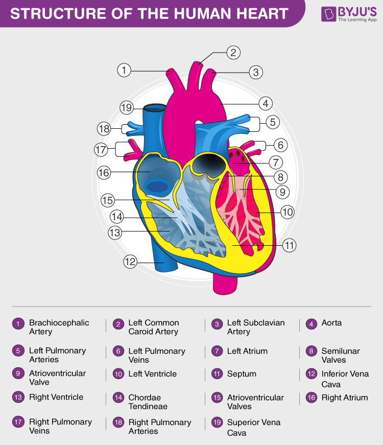
The human being heart is near the size of a human fist and is divided into four chambers, namely two ventricles and 2 atria. The ventricles are the chambers that pump blood and atrium are the chambers that receive blood. Amid which both right atrium and ventricle make up the "right heart," and the left atrium and ventricle make up the "left heart." The structure of the heart too houses the biggest artery in the trunk – the aorta.

The right and the left region of the heart are separated by a wall of muscle chosen the septum. The right ventricle pumps the blood to the lungs for re-oxygenation through the pulmonary arteries. The right semilunar valves shut and preclude the blood from flowing back into the eye. Then, the oxygenated blood is received past the left atrium from the lungs via the pulmonary veins. Read on to explore more about the structure of the eye.
External Structure of Center
One of the very first structures which can be observed when the external structure of the center is viewed is the pericardium.
Pericardium
The man eye is situated to the left of the chest and is enclosed within a fluid-filled crenel described every bit the pericardial cavity. The walls and lining of the pericardial cavity are made up of a membrane known as the pericardium.
The pericardium is a fibre membrane found as an external covering around the center. It protects the heart by producing a serous fluid, which serves to lubricate the heart and prevent friction between the surrounding organs. Apart from the lubrication, the pericardium too helps by holding the heart in its position and by maintaining a hollow space for the center to aggrandize itself when it is full. The pericardium has two sectional layers—
- Visceral Layer:Information technology directly covers the outside of the heart.
- Parietal Layer: Information technologyforms a sac around the outer region of the heart that contains the fluid in the pericardial cavity.
Structure of the Heart Wall
The heart wall is made up of 3 layers, namely:
- Epicardium – Epicardium is the outermost layer of the heart. Information technology is composed of a sparse-layered membrane that serves to lubricate and protect the outer section.
- Myocardium – This is a layer of muscle tissue and information technology constitutes the centre layer wall of the heart. Information technology contributes to the thickness and is responsible for the pumping action.
- Endocardium – It is the innermost layer that lines the inner middle chambers and covers the heart valves. Furthermore, it prevents the blood from sticking to the inner walls, thereby preventing potentially fatal blood clots.
Internal Structure of Centre
The internal structure of the heart is rather intricate with several chambers and valves that command the flow of blood.
Chambers of the Heart
Vertebrate hearts can be classified based on the number of chambers present. For case, most fish have two chambers, reptiles and amphibians have three chambers. Avian and mammalian hearts consists of 4 chambers. Humans are mammals; hence, we have four chambers, namely:
- Left atrium
- Right atrium
- Left ventricle
- Right ventricle
Atria are thin, less muscular walls and smaller than ventricles. These are the claret-receiving chambers that are fed by the large veins.
Ventricles are larger and more muscular chambers responsible for pumping and pushing blood out to the circulation. These are connected to larger arteries that deliver blood for circulation.
The right ventricle and right atrium are insufficiently smaller than the left chambers. The walls consist of fewer muscles compared to the left portion, and the size deviation is based on their functions. The blood originating from the right side flows through the pulmonary circulation, while blood arising from the left chambers is pumped throughout the body.
Blood Vessels
In organisms with airtight circulatory systems, the blood flows inside vessels of varying sizes. All vertebrates, including humans, possess this type of circulation. The external construction of the eye has many blood vessels that form a network, with other major vessels emerging from within the construction. The blood vessels typically comprise the post-obit:
- Veins supply deoxygenated blood to the eye via inferior and superior vena cava, and information technology somewhen drains into the right atrium.
- Capillaries are tiny, tube-like vessels which course a network between the arteries to veins.
- Arteries are muscular-walled tubes mainly involved in supplying oxygenated blood away from the heart to all other parts of the body. Aorta is the largest of the arteries and it branches off into various smaller arteries throughout the body.
Also Refer: Difference between Arteries and Veins
Valves
Valves are flaps of fibrous tissues located in the cardiac chambers between the veins. They ensure that the blood flows in a single direction (unidirectional). Flaps also prevent the claret from flowing backwards. Based on their function, valves are of two types:
- Atrioventricular valves are between ventricles and atria. The valve between the right ventricle and right atrium is the tricuspid valve, and the i which is constitute between the left ventricle and left atrium is known as the mitral valve.
- Semilunar valves are located between the left ventricle and aorta. It is also found between the pulmonary artery and right ventricle.
Also Read: Blood and its Composition
Facts about Human Center

- The center pumps around 5.vii litres of blood in a day throughout the torso.
- The center is situated at the centre of the chest and points slightly towards the left.
- On average, the middle beats most 100,000 times a 24-hour interval, i.e., around 3 billion beats in a lifetime.
- The average male person center weighs around 280 to 340 grams (10 to 12 ounces). In females, it weighs around 230 to 280 grams (8 to 10 ounces).
- An adult heart beats most lx to 100 times per minute, and newborn babies heart beats at a faster pace than an developed which is well-nigh 90 to 190 beats per minute.
As well Refer:Center Health
To know more nigh the human heart structure and role, or whatever other related concepts such as arteries and veins, the internal construction of heart, the external construction of middle, due eastxplore at BYJU'S Biology. Also, discovereasy diagram of heart, concepts and relevant questions for human being heart for Class x by downloading BYJU'S – The Learning App.
More to Explore:
- Hypoxia
- Heart Diseases
- Hepatic Portal System
Oft Asked Questions
1. What is pulmonary circulation? Explicate.
Pulmonary circulation is a blazon of claret circulation responsible for conveying deoxygenated blood away from the heart, and to the lungs, where it is oxygenated. The system and so brings oxygenated claret back to the heart to exist pumped throughout the body.
ii. Define systemic circulation.
In the systemic apportionment, the heart pumps the oxygenated blood through the arteries to every organ and tissue in the body, and then dorsum again to the heart through a system of veins.
three. Elaborate coronary apportionment and its significance.
The eye is a musculus, and it needs a abiding supply of oxygenated claret to survive and piece of work effectively. This is where coronary circulation fulfils this function through a network of arteries and veins in the middle. The coronary arteries supply oxygenated blood to the eye, and the cardiac veins bleed the blood in one case it has been deoxygenated by the tissues of the centre.
4. Briefly explain the construction of the man heart.
The homo heart is divided into four chambers, namely two ventricles and two atria. The ventricles are the chambers that pump blood and atrium are the chambers that receive the blood. Among which, the right atrium and ventricle make up the "correct portion of the eye", and the left atrium and ventricle make upward the "left portion of the center."
5. Proper noun the chambers of the middle.
- Left atrium
- Right atrium
- Left ventricle
- Right ventricle
six. What is pericardium? Explain its role.
The pericardium is a fibrous membrane that envelops the heart. It too serves a protective part by producing a serous fluid, which lubricates the heart and prevents friction betwixt the surrounding organs. Furthermore, the pericardium also holds the heart in its position and provides a hollow infinite for the heart to expand and contract.
7. Explain the three layers of the heart wall.
The middle wall is made up of 3 layers, namely:
- Epicardium – This is the outermost layer of the heart. It is equanimous of a sparse layer of membrane that protects and lubricates the outer department.
- Myocardium – This is a layer of muscle tissue that constitutes the center layer wall of the heart. It is responsible for the centre'south "pumping" activeness.
- Endocardium – The innermost layer that lines the inner heart chambers and covers the heart valves. Prevents blood from sticking, thereby avoiding the formation of fatal claret clots.
8. Explicate the 3 major blood vessels of the human torso.
The claret vessels comprise:
- Veins – Information technology supplies deoxygenated claret to the heart via inferior and superior vena cava, eventually draining into the correct atrium.
- Capillaries – They are minuscule, tube-similar vessels which class a network between the arteries and veins.
- Arteries – These are muscular-walled tubes responsible for supplying oxygenated claret away from the eye to all other parts of the body.
ix. What is the function of the middle valves? Provide examples of various valves.
Valves are flaps of tissues that are present in cardiac chambers between the veins. They forestall the backflow of blood. Examples include the atrioventricular valves, tricuspid valves, mitral valves and the semilunar valves.
10. What is meant by myocardial infarction?
Myocardial infarction is a serious medical status where the claret period to the eye is reduced or entirely stopped. This causes oxygen deprivation in the middle muscles, and prolonged deprivation tin can cause tissues to die.
Source: https://byjus.com/biology/human-heart/
0 Response to "Can We Stop Human Heart and Start It Again"
Post a Comment